
The infra-red elegans of worms feeding on carbon nanotubes
The infra-red elegans of worms feeding on carbon nanotubes
Eddie Sharaga, who recently graduated with his Master’s degree under the supervision of Prof. Gili Bisker, found himself unprepared for the encounter with the notorious Caenorhabditis Elegans (C. elegans) worms. It turned out to be a close encounter, almost microscopic in nature, one might say.
In their recently published paper in Advanced Materials Technologies, titled “Spatiotemporal Tracking of Near-Infrared Fluorescent Single-Walled Carbon Nanotubes in C. Elegans Nematodes Confined in a Microfluidics Platform“, Eddie describes a novel infra-red in vivo imaging platform for C. elegans nematodes utilizing single-walled carbon nanotubes (SWCNTs) as optical sensors.
Prof. Bisker and her team are engaged in researching optical nanosensors utilizing the distinctive optical properties of SWCNTs for precise and selective molecular recognition in biological systems. Recent investigations in her laboratory have also examined the application of SWCNTs as optical markers for in vivo imaging of model organisms, such as the C. elegans nematode. Furthermore, Bisker investigates complex out-of-equilibrium self-assembly inspired by biological systems.
Single Walled Carbon nanotubes as in vivo imaging probes in C. elegans nematodes
C. elegans nematodes are a powerful model organism for diverse biological and biomedical studies, benefiting from their genetic similarities to humans, small size, and transparency. However, live fluorescence imaging of C. elegans can be challenging due to the strong autofluorescence in the visible range, which obscures the signal of common fluorescent proteins or dyes. SWCNTs fluoresce in the near-infrared (NIR) range, where there is no autofluorescence background.
In this research, Eddie developed a platform for in vivo NIR imaging of C. elegans gastrointestinal tract using biocompatible SWCNTs. The SWCNTs serve as fluorescent tracking probes within the worm gut, following internalization along with food intake. A microfluidic confinement device was employed to ensure an anesthetics-free feeding environment, allowing spatiotemporal control over the SWCNTs intake imaging.

Figure from the paper: Worm confinement effect on colocalization mismatch of autofluorescence and NIR channels. a) 3D explosion of separated brightfield, NIR fluorescence, and visible fluorescence multichannel layering images. b) Overlay image of a free-roaming worm with SWCNTs showing colocalization challenge. c) Overlay image of a confined worm with SWCNTs.
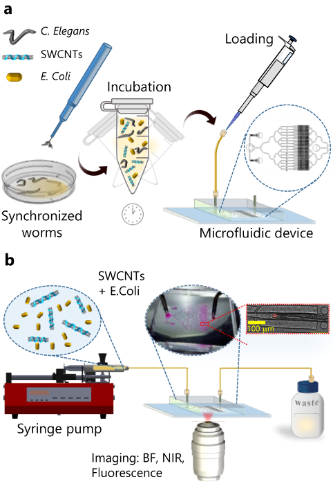
Schematic illustration of the experimental setup and procedures. a) C. elegans are loaded into the microfluidic platform. b) Real-time fluorescence imaging of the SWCNTs within the worms that are confined in the microfluidic device.
 What is new?
In this paper, an improved colocalization of spectrally separated fluorescence images was demonstrated, along with real-time imaging of SWCNTs optical nano-probes used for digestive trajectory tracking.
What is the contribution to future research?
Owing to the unique optical properties of SWCNTs and the confinement of the worms in the microfluidics system, the proposed platform facilitates advanced in vivo imaging of C. elegans in both the visible and NIR regions, opening numerous avenues for advancing research of C. elegans and other microscopic model organisms.